Updated by Piper Wenzel, BS May 2024 and Samantha Corman, BS, MA September 2026
See also:
Background
- Also known as 2nd branchial cleft remnant or branchial cleft anomaly (Holt 2025)
- Congenital developmental defect in 2nd branchial apparatus. Most accepted theory of pathogenesis is a failure of complete obliteration of branchial cleft. May manifest as a sinus, fistula, cyst, or any combination of the three (Bajaj 2011, Mehmi 2019, Holt 2025)
- The most common branchial cleft anomaly, making up 40-95% of all branchial cleft anomalies (Mehmi 2019, Holt 2025)
- Can occur anywhere along the tract of the branchial cleft, from the tonsillar fossa to the supraclavicular region. The characteristic location of a sinus fistula ostium is anteromedial to the sternocleidomastoid (Holt 2025)
- May be in proximity to nerves (glossopharyngeal or hypoglossal) and carotid vessels (Holt 2025)
- Bilateral second branchial cleft cysts may be associated with branchio-oto-renal syndrome (Holt 2025)
- Upper respiratory tract infections can result in "increase in size of swelling, purulent discharge and abscess formation" (Mehmi 2019)
Classification System for 2nd Branchial Cleft Anomaly (Bailey 1929)
- Type I: deep to platysma and cervical fascia
- Type II: adjacent to carotid sheath
- Type III: between internal carotid artery and external carotid artery
- Type IV: medial to carotid sheath, adjacent to lateral pharyngeal wall
Radiological Findings
- On CT:
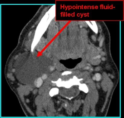
- Low density, unilocular on non-contrast, may have septations if infected
- Low density with non-enhancing wall on contrast CT, infection can appear as multilocular structure with fat-standing and wall-enhancement
- On MR:
- T1 shows fluid signal isointense to CSF, infected cysts may have hyperintense proteinaceous contents
- T2 is hyperintense
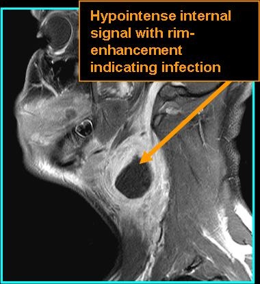
- T1 post-contrast shows no enhancement, unless infection is present in which case the cyst may show ring-enhancement



References
Bailey H. Branchial cysts and other essays on surgical subjects in the facio‑cervical region. London: Lewis, 1929. viii, 86 p. ill. https://doi.org/10.1002/bjs.1800176648
Bajaj Y, Ifeacho S, Tweedie D, et al. Branchial anomalies in children. Int J Pediatr Otorhinolaryngol. 2011;75(8):1020-1023. doi:10.1016/j.ijporl.2011.05.008
Holt AC, Lofgren DH, Hohman MH, Shermetaro C. Branchial Cleft Anomalies. In: StatPearls. Treasure Island (FL): StatPearls Publishing; May 27, 2025.
Mehmi N, Kumar R, Sagar P, et al. Importance and Impact of Appropriate Radiology in the Management of Branchial Cleft Anomalies. Indian J Otolaryngol Head Neck Surg. 2019;71(Suppl 1):953-959. doi:10.1007/s12070-019-01634-w