Return to: Sialograms and Sialography
Case History and Images
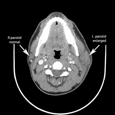
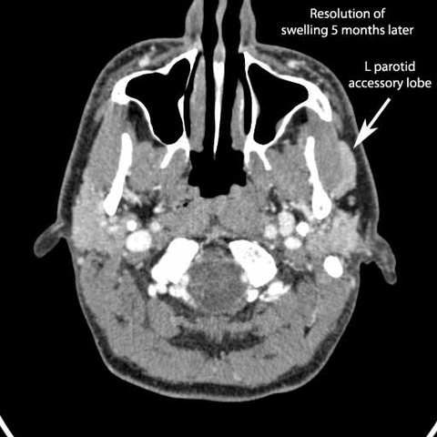
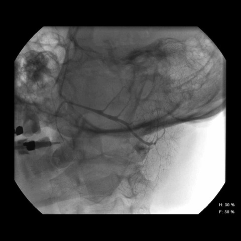
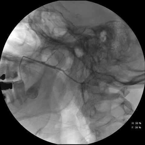
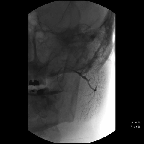
Patient with two episodes of parotid swelling with initial CT (without contrast) showing swelling, second CT showing only accessory lobe. Subsequent sialogram normal.
|
|
| 
|
Return to: Sialograms and Sialography
Patient with two episodes of parotid swelling with initial CT (without contrast) showing swelling, second CT showing only accessory lobe. Subsequent sialogram normal.


|

|

| 
|