Return to: Sentinel Lymph Node Biopsy (contains information not only about false negatives, but also controversy regarding the use of the technique)
See also:
- Posterolateral Neck Dissection
- Case Example Posterolateral Neck Dissection
- Case example 2 Posterolateral Neck Dissection and Anatomy
Video
Clinical Images
|
|
|
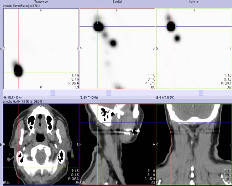
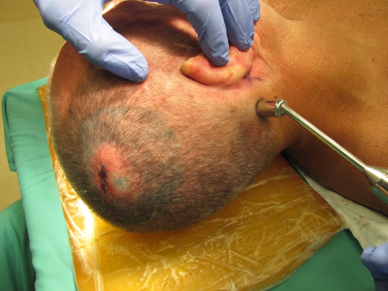
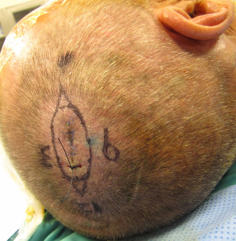
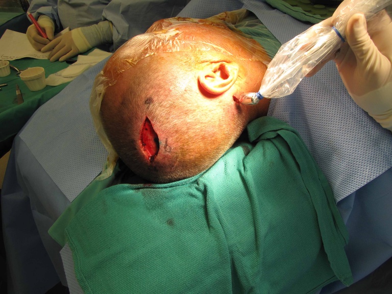
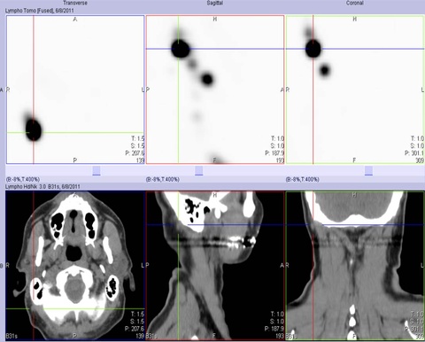
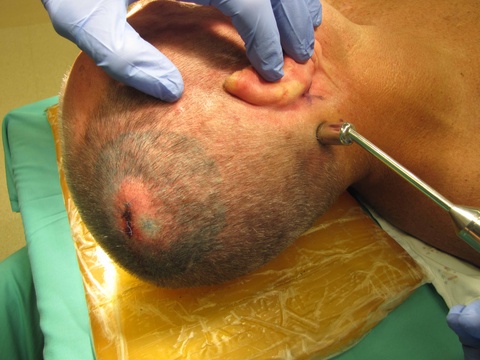
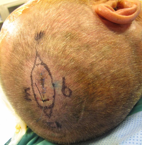
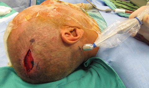
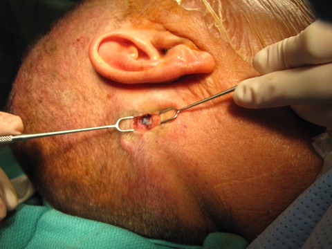
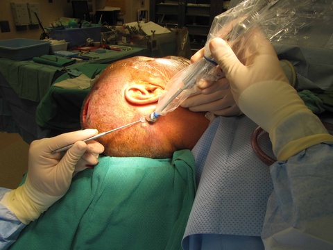
| Lymphoscintigram with CT co-registration. Note the post-auricular/anterior occipital node identified within the cross-hairs on the three CT images. | Gamma probe identifying activity from the Tc99 injection performed 3 hours previously for lymphoscintigram. | 1 cm margins about T1b melanoma oriented with suture at 12:00. Note region (at 9:00) of 0.5 cc of dermal injection of methylene blue. |
|
|
|
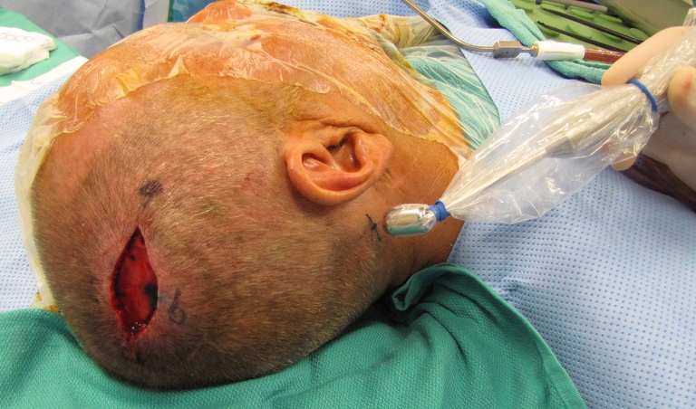
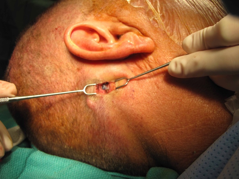
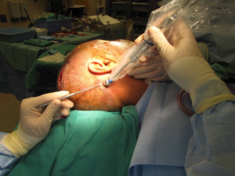
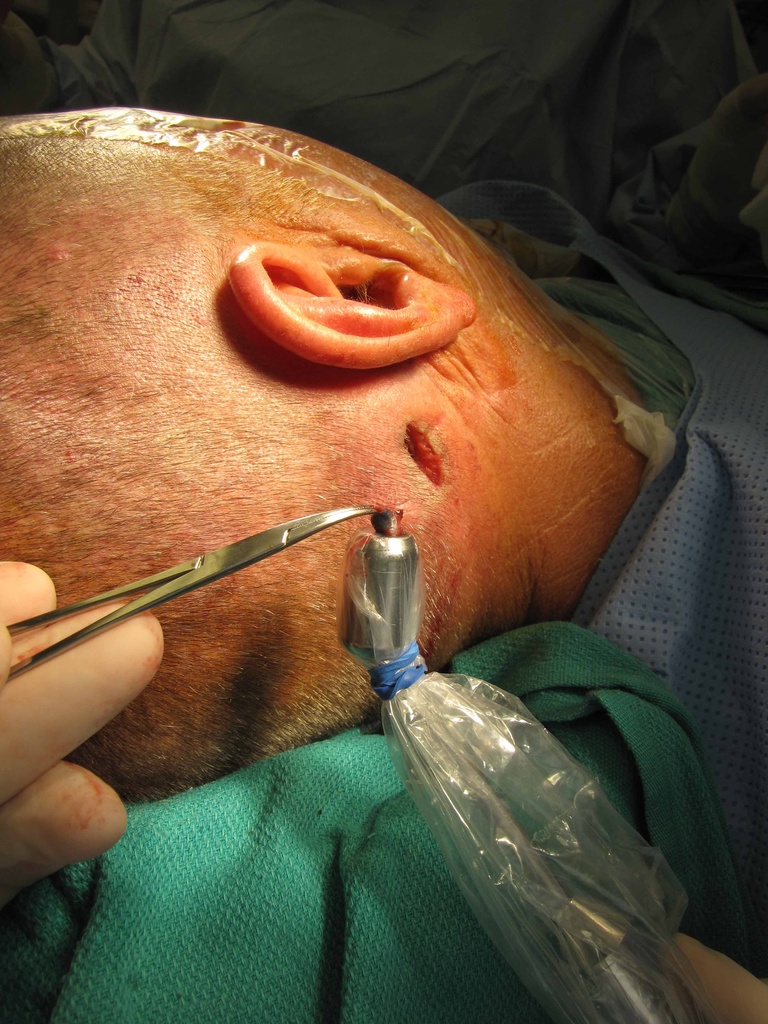
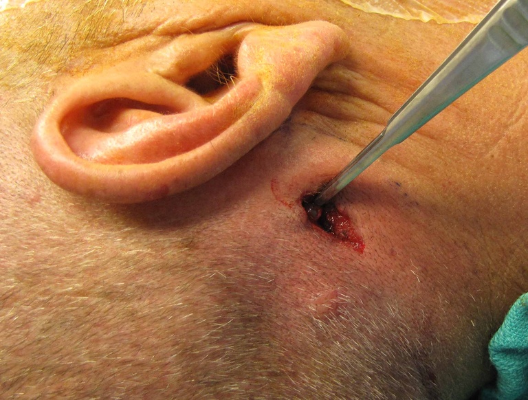
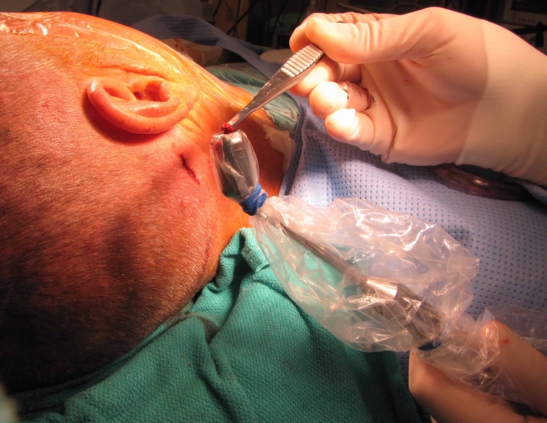
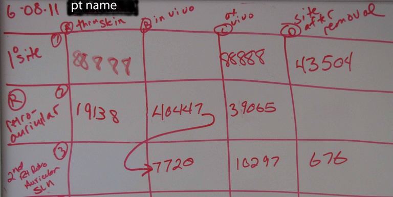
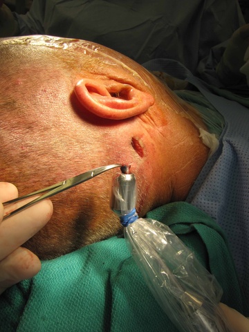
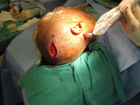
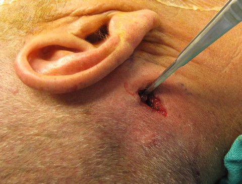
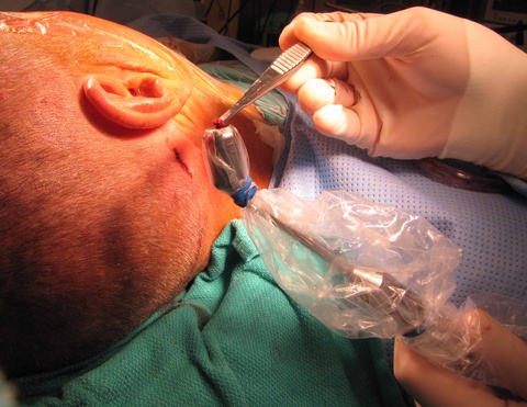
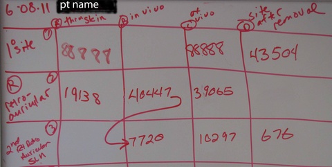
| Resection of primary site is done first - with planning for incision for SLN bx directed by gamma probe and consideration for potential need for subsequent surgery if node positive (postero-lateral neck dissection) | Superficial suboccipital node is identified with gamma probe with blue color (from methylene blue injection) further helping identification. | "In vivo" 10 second count with gamma probe is recorded as 40,447 on grid (see last photo). "In vivo" refers to evaluation with the node exposed through the incision. |
|
|
|
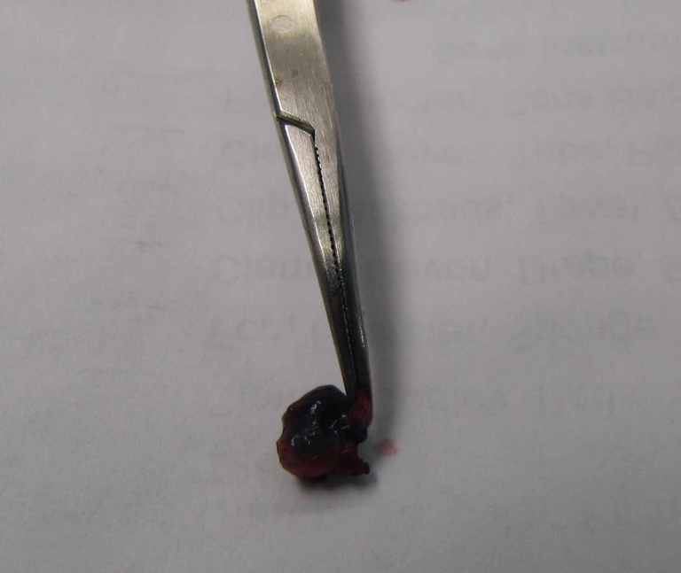
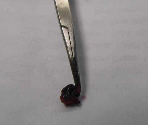
| "Ex vivo" analysis with the node removed is recorded as 39,065. | Appearance of resected blue node. Note that grasping soft tissue adjacent the node (and not the node itself) facilitates traction during removal. | "Site after removal" is recorded as 7,720 - still too high to confirm removal of SLN (7,720/40,447 x 100 = 19.1%) - a drop to 10% activity is required. |
|
|
|
| Further dissection directed by the gamma probe activity reveals an adjacent, deeper blue node removed and tested. | Activity in the second node ("ex vivo") is 10,297. | Final analysis of "site after removal" activity of 676 identifies a decrease from 40,447 (676/40,447 x 100 =1.7% activity) - confirming removal of sentinel nodes. |
|
|
|
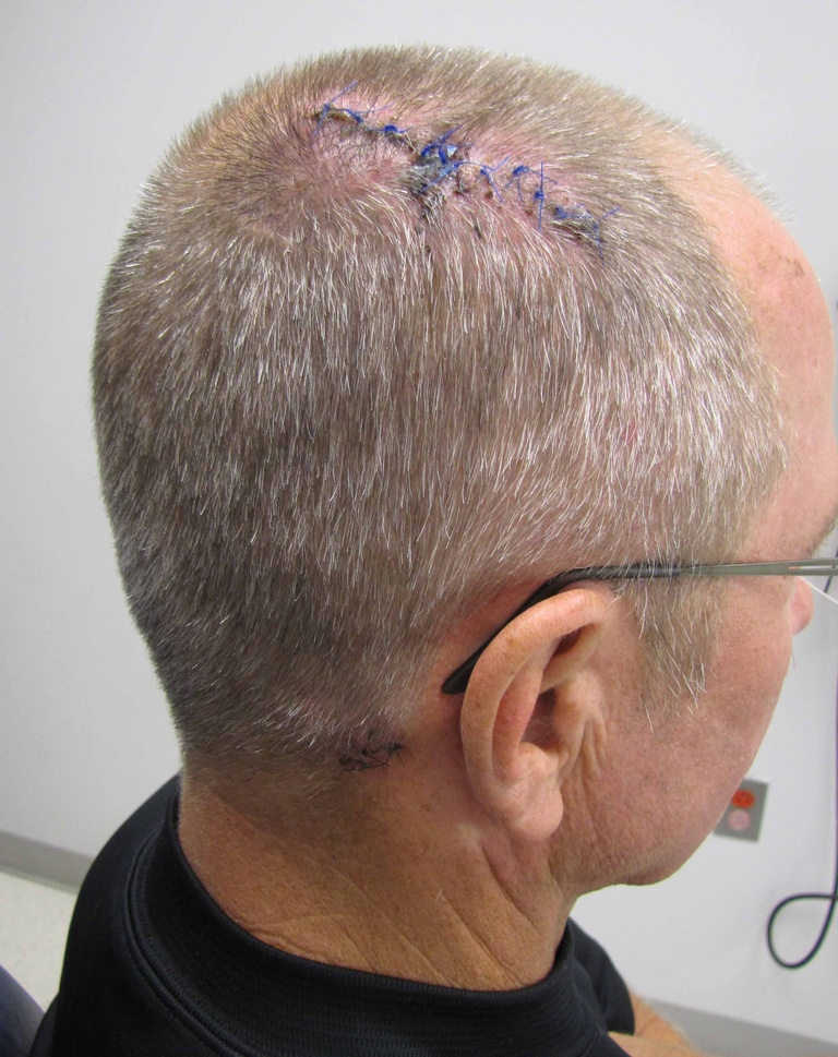
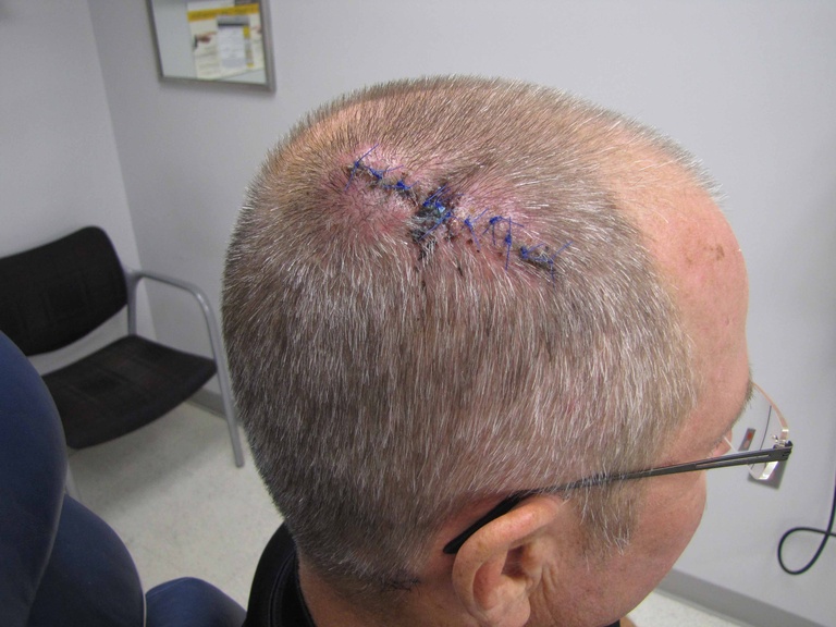
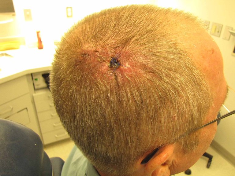
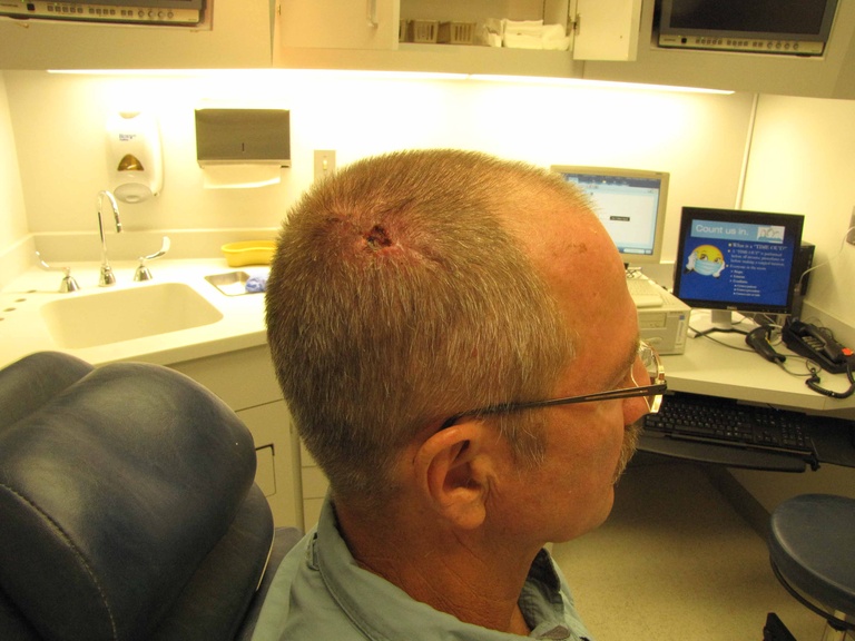
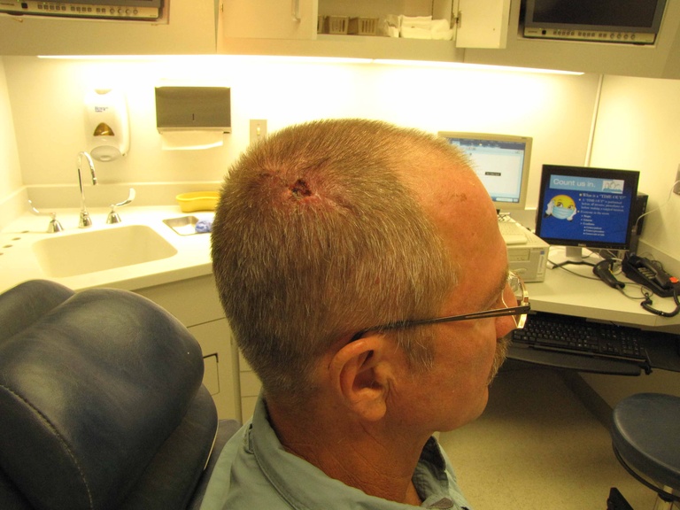
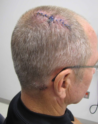
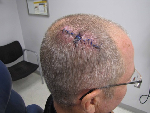
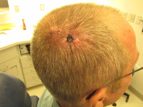
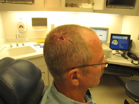
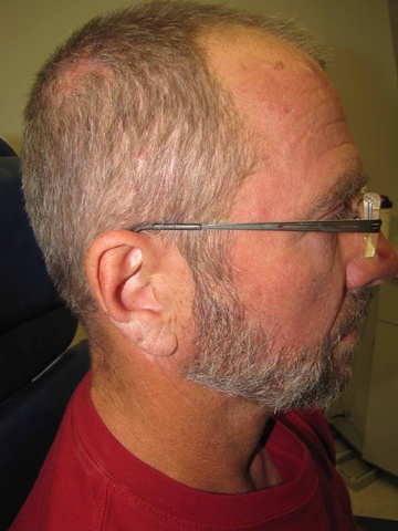
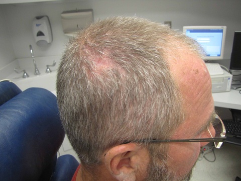
| followup one week postop | followup one week postop | followup two weeks postop |
|
|
|
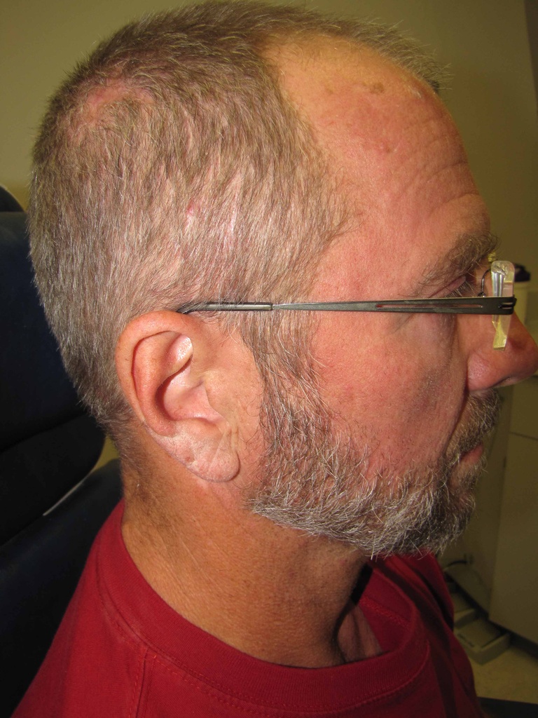
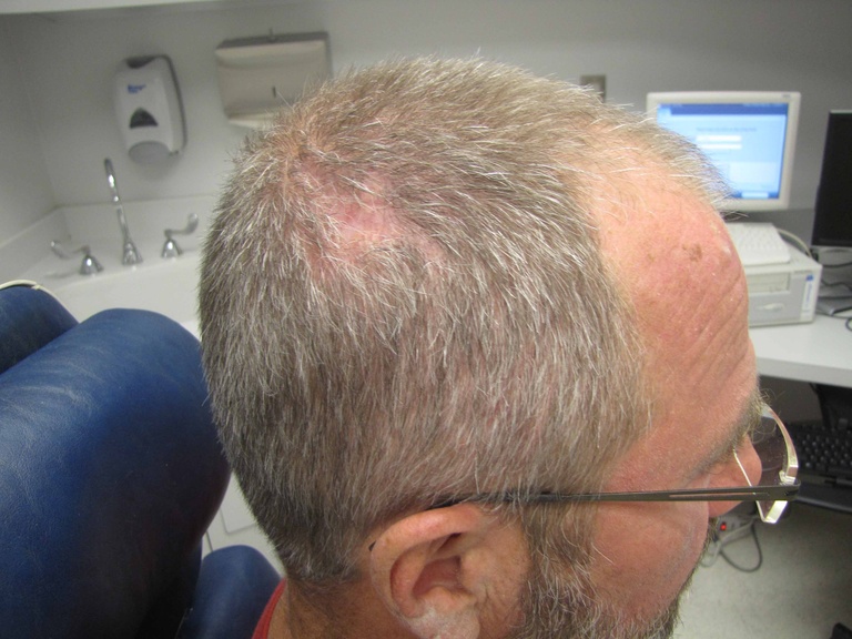
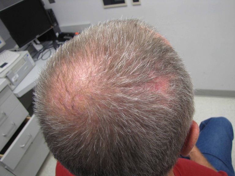
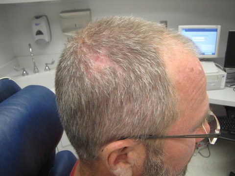
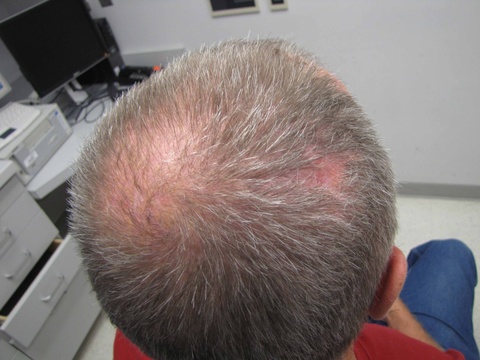
| followup two weeks postop | followup 3 months postop | followup 3 months postop |
|
| |
| followup 3 months postop | followup 3 months postop |
