Return to protocol:
See also:
Case History
41 yo female with right submandibular gland swelling occuring several times per week, usually with dinner, lasting for an hour followed by resolution.
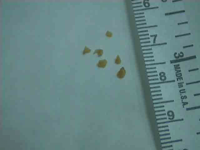
Imaging: CT scan = 3 mm calicified calculus (Sialolithiasis) in upper margin of right submandibular gland with the gland otherwise unremarkable
Sialendosocpy of right SMG done under general anesthesia
First Video
- Canulation of duct with 0.018 inch guide
- Dilation of duct orifice
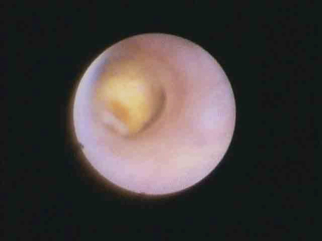
- Following guide with 1.6 (o.d.) Zenk sialendoscopy (video begins)
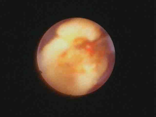
- Laser fragmentation of stone (holmium laser on)
- Basket removal of stone
Second Video
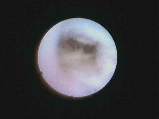
- Removal of multiple other fragments (basket and forceps)
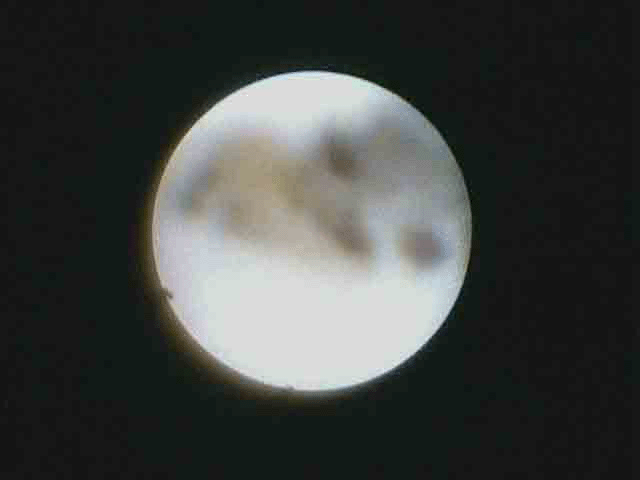
- View of cleared hilum
