return to: Sialography - How To and Do I Need To? Contemporary Infusion Digital Sialography
Case #2
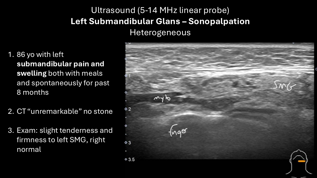
Presentation: 86 yo with left submandibular pain and swelling both with meals and spontaneously over 8 months.
Exam: slight tenderness and firmness to left submandibular gland, right is normal
CT: unremarkable salivary glands - no stone
Ultrasound (POCUS at time of visit):
Panel Discussion Regarding Management
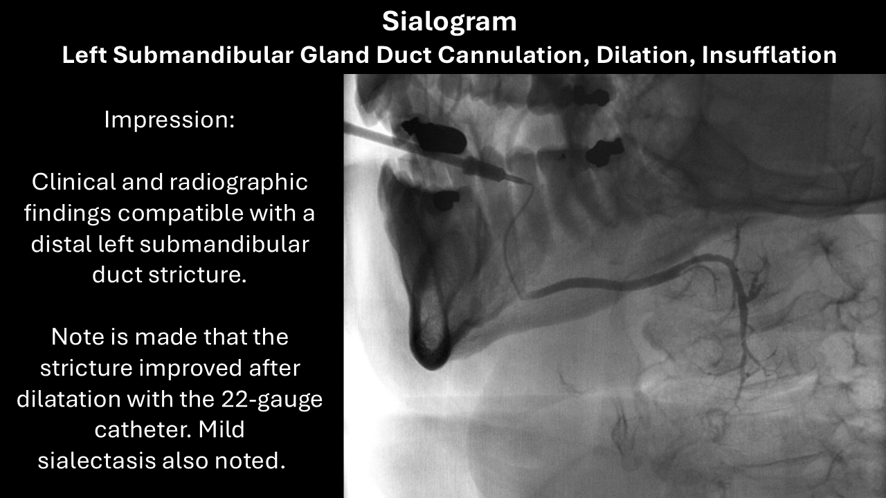
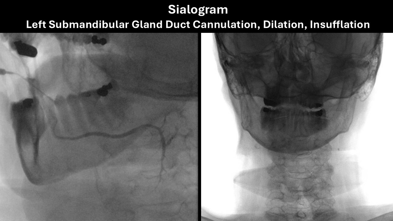
Sialogram:
Outcome:
- Immediately after sialogram in clinic: "this was now the best her left submandibular gland had felt in 2 years". Exam: Successful massage of the gland with the duct showing clear material (saliva vs Isovue®) emanating from it. We suggested maintenance of good hydration and massage would be helpful to hopefully prevent recurrence with the understanding which were she to have recurrence more aggressive dilation could be done possibly in the clinic
- Symptom Free for 9 months then recurrence of symptoms
- In Clinic Procedure Duct Dilation 10 months after sialogram - (see further below)
- Symptom Free at Last Follow-up (10 months after in clinic dilation; 20 months after sialogram)
Procedure Note (modified)
In-Clinic Dilation (10 months after sialogram)
Left submandibular duct cannulation dilation with Kenalog® 10 diluted with lidocaine insufflation (2.5 cc of "Kenalog® 10" created by mixing Kenalog 40 part with 3 parts of 1% lidocaine)
Preop Diagnosis: Left submandibular sialoadenitis with ductal narrowing previously responsive to a sialogram
Postop Diagnosis: Same
Anesthesia: Premedication with antibiotics as well as oral application of 2% viscous lidocaine and Betadine swab to the floor the mouth
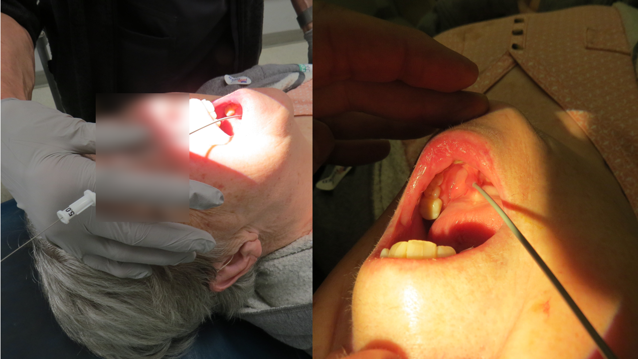
Description of Procedure: Following identification, patient informed consent, and a brief timeout, patient was placed in the semi recumbent fashion. A microscope was brought in from above. There was initial difficulty in identifying the left submandibular duct orifice, which was facilitated by massage of the right to identify the contralateral equivalent duct orifice. Attention was then brought back to the left side with ipsilateral massage allowing for placement of a Rosen needle (Turner needle) followed by manipulations with 2 DeBakey forceps to straighten the duct. Cannulation was achieved with a 0.015 inch guidewire 15 cm long. Using Seldinger technique, dilation was achieved over the wire with a 24-gauge Angiocath followed by a 22-gauge Angiocath.
With the 22-gauge angiocath in place, the guidewire was removed and "Kenalog 10®" ["Kenalog 10®" = one part Kenalog 40®, three parts 1% lidocaine] preparation was slowly instilled to a volume of 2.5 cc. Patient affirmed that the pain was similar in nature, though decreased in severity to that which she has had with eating. After the initial pain on insufflation, she had no further pain and even reported relief of the pain from that which she came in with.
A 30 cm long 0.015-inch guidewire was placed through the 22-gauge Angiocath. The Angiocath was then removed. Further dilation was then achieved successively using a 5, 6 then final 7 French Cook salivary access dilator placed to a depth of approximately 3 cm. The French Cook salivary access dilators were successfully left in place for 2 minutes for the 5 and 1 minute for the 7.
She tolerated the procedure well with no pain on inspection of the area and photographs taken during the course of the procedure.