Goals
- Standardize salivary ultrasound nomenclature and technique with consistency reporting of the specific subsites within the glands
- Additional benefit: provide consistency within the evolving field of ultrasound radiomics (elastography)
- Acknowledge shortcomings including “the salivary glands are dynamic structures whose blood flow, secretory status, size and location have been reported to be modified by circadian rhythms, degree of hydration…” and other (for example – salivary stimulation with ascorbic acid etc)
- Define subunits of the gland
Subsites |
Superficial lobe |
| Buccal extension |
-Facial Process |
Lateral to mandible Accessory Lobe Facial Process |
| Posterior to masseter |
| Tail of parotid |
Deep lobe |
| Medial to mandible |
- Define optimal measurement for size with use of terms “Length” “Height” and “Depth”
- Discuss technique (with likely affirmation from the AIUM
- Use of linear probe - ~ 5-14 MHz
- Acknowledge impact of pressure of probe on salivary dimensions (including shear wave)-
- Define terminology to identify more specific sites within the submandibular gland
- "Subunits” = Portions of gland described as: Midportion / Anterior / Posterior / Superficial / Deep / Superior / Inferior
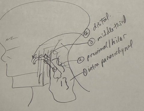
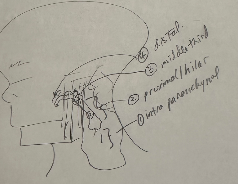
- Affirm pre-existing classification system for stone location
- Classification of stone location (Goncalves et al 2017)
- Sialolithiasis Definition: "Hyperechoic reflexes with distal signal loss along the course of the duct"
- From Goncalves et al "Sonography in Diagnosis of Sialolithiasis" J Ultrasound Med 2017;36:2227-2235
- Sialolithiasis Definition: "Hyperechoic reflexes with distal signal loss along the course of the duct"

- Parotid sonographic landmarks
- Intraparenchymal stone proximally located in parenchyma
- Proximal/hilar stone 1 cm proximal to the posterior edge of masseter to middle of masseter muscle
- Middle third middle of masseter muscle to to anterior edge of masseter
- Distal duct including papillary region anterior edge of masseter muscle to papilla
- To refine the language we use in analyzing, treating and discussing the salivary glands
Multi-year Plan to Establish Standardization
- Part 1 Submandibular Gland
- Part 2 Parotid Gland
- Part 3 Sublingual and Minor Salivary Glands
- Part 4 Radiomics: Shear Wave Elastography
