Return to: Parotidectomy with Facial Nerve Dissection
See also:
- Case example - neck apocrine cystadenoma presenting clinically similar to lipoma
- Lipoma Radiology; Liposarcoma Radiology
Follow-Up Images
10 years after surgery.
Initial Clinical Presentation
Patient presented in November 2008 with "salivary swelling" to the left parotid gland. Reported that the swelling "comes and goes" and "has been somewhat responsive" to antibiotics and conservative measures such as massage, but it "never completely goes away."
January 2009 CT: Well circumscribed fatty dense lesion in the left parotid gland. This could represent lipomatous tumor such as lipoma.
Preoperative MRI: Left parotid lipomatous tumor, containing small enhancing lesion with differential of vascular structure versus solid lesion. Repeat ultrasound to further define the possible vasculature is recommended.
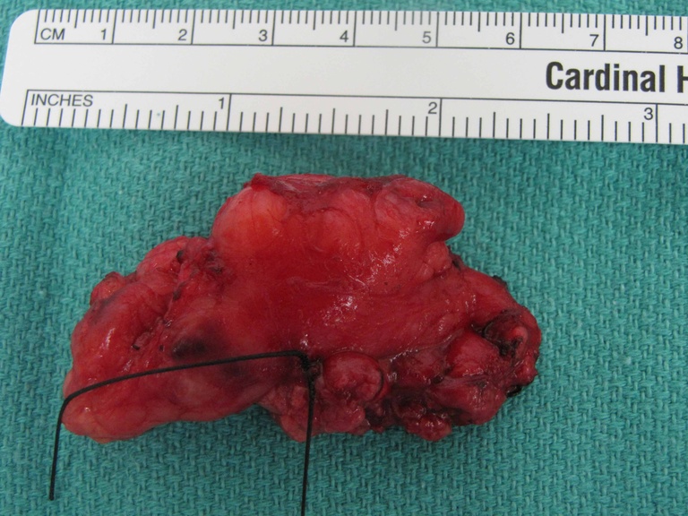
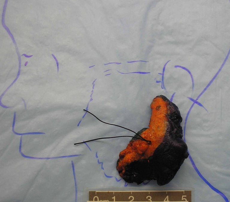
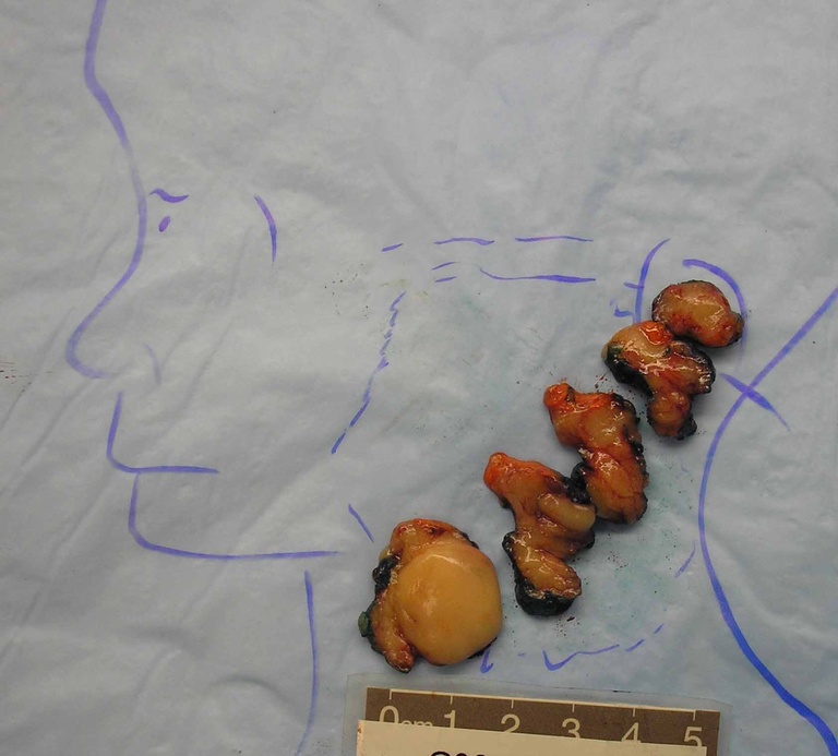
Diagnosis: (pathology report after first resection) Atypical lipomatous neoplasm, favor well differentiated, lipoma-like liposarcoma, 2.3 cm in greatest dimension, abutting the lateral surgical resection margin.
Surgical Management
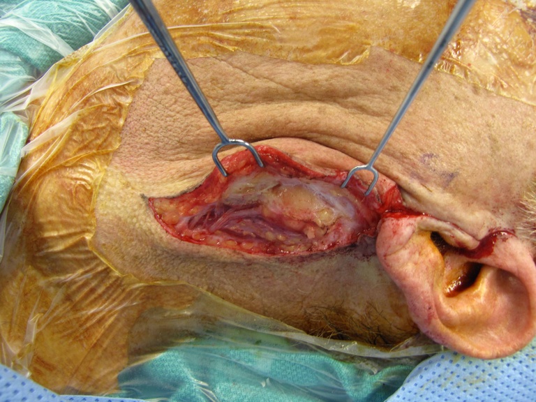
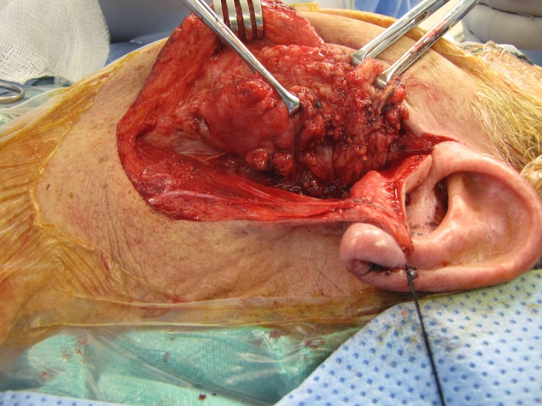
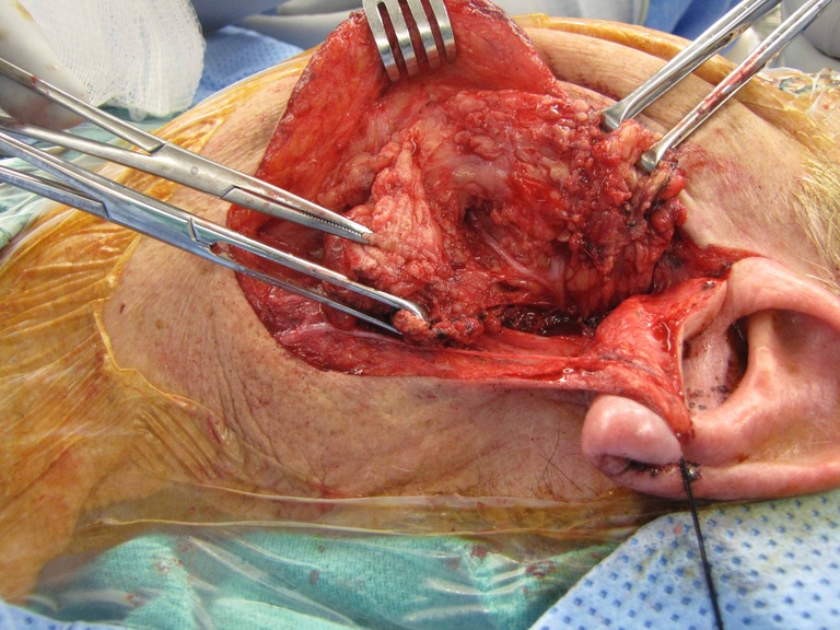
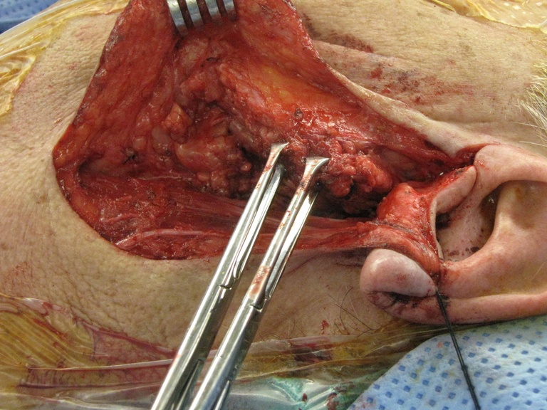
1st Resection - Parotidectomy with Facial Nerve Dissection and Preservation of the Great Auricular Nerve
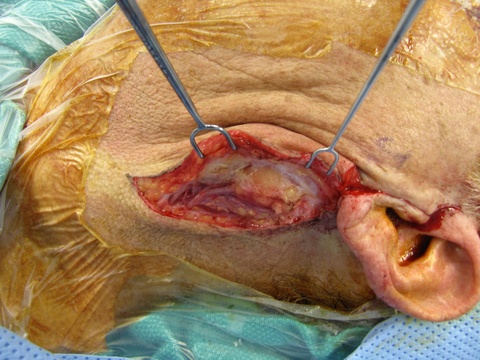
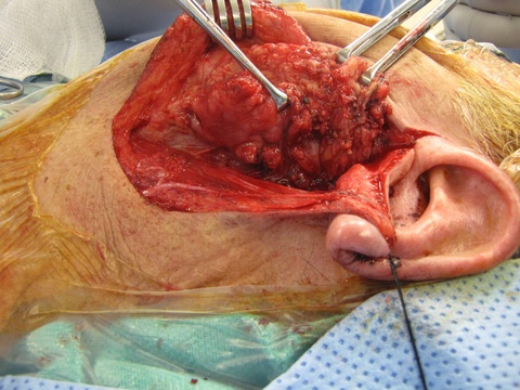
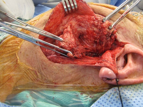
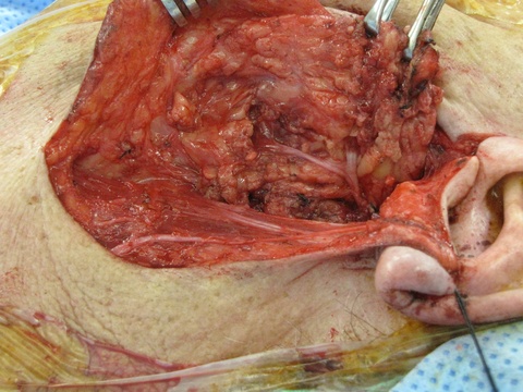
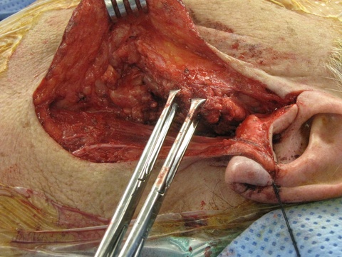
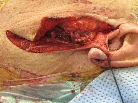
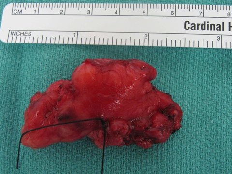
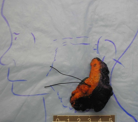
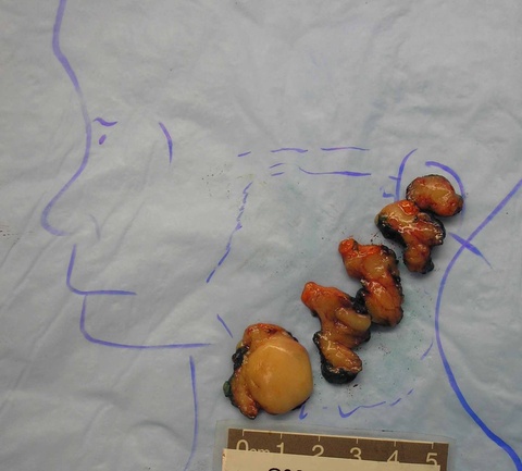
Pathology comment: This lipomatous tumor shows nuclear pleomorphism and irregularity, and given the sub-fascial location, the tumor is best interpreted as lipoma-like liposarcoma. This low-grade tumor may have a small risk of local reoccurrence given the close lateral margin. Spindle cell lipoma was also a consideration, however the CD34 stain is negative.
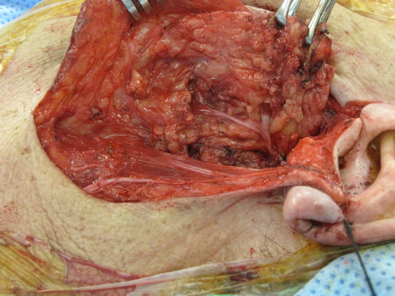
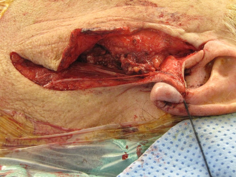
2nd Resection - 7 Days Later to Clear Positive Margins
Requiring sacrifice of the great auricular nerve
